
Anti-p34 / cdk1(CDK1/873)
CAT:
37-BNUB0873-500
Size:
500 µL
Price:
Ask
- Availability: 24/48H Stock Items & 2 to 6 Weeks non Stock Items.
- Dry Ice Shipment: No


Anti-p34 / cdk1(CDK1/873)
- Description: Recognizes a 34 kDa protein, which is identified as cyclin dependent kinase 1 (cdk1) or p34cdc2 protein kinase. cdk1 / p34cdc2 plays a crucial role during cell division and is most active during mitosis. It is predominantly localized in the nucleus. It is a serine/threonine kinase, which is activated by cyclin, presumably by de-phosphorylation of tyrosine residues. Activated cdk1 / p34cdc2 performs specific functions during mitosis, including nuclear envelope breakdown and chromosome condensation.Primary antibodies are available purified, or with a selection of fluorescent CF® Dyes and other labels. CF® Dyes offer exceptional brightness and photostability. Note: Conjugates of blue fluorescent dyes like CF®405S and CF®405M are not recommended for detecting low abundance targets, because blue dyes have lower fluorescence and can give higher non-specific background than other dye colors.
- Synonyms: Cdc2; CDC28A; CDK1; CDKN1; CELL CYCLE CONTROLLER CDC2; Cell division control protein 2 homolog; Cell division cycle 2 G1 to S and G2 to M; Cell division protein kinase 1; Cyclin-dependent kinase 1; p34 Protein Kinase; P34CDC2
- CAS Number: 9007-83-4
- UNSPSC: 41116161
- UNSPSC Description: Primary and secondary antibodies for multiple methodology immunostaining detection application
- Gene Name: CDK1
- Gene ID: 983
- NCBI Gene ID: 334562
- UniProt: P06493
- Cellular Locus: Nucleus & cytoplasm
- Host: Mouse
- Species Reactivity: Cow, Human, Mink, Monkey
- Immunogen: Recombinant human CDK1 protein
- Target Antigen: Cdk1 | p34
- Clonality: Monoclonal
- Isotype: IgG2a κ
- Clone: CDK1/873
- Conjugation: Purified, with BSA
- Source: Animal
- Applications: IHC, FFPE (verified) | WB (verified)
- Validated Applications: IHC, FFPE, WB
- Field of Research: Cell cycle, Tumor suppressors
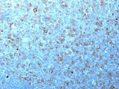
- Positive Control: HeLa Cells or Tonsil
- Concentration: 0.2 mg/mL
- Buffer: PBS, 0.05% BSA, 0.05% azide
- Molecular Weight: 34 kDa
- Additionnal Information: Higher concentration may be required for direct detection using primary antibody conjugates than for indirect detection with secondary antibody|Immunofluorescence: 1-2 ug/mL|Does not react with mouse, rat, Xenopus, Drosophila, S. pombe, or S. cerevisiae; others not known|Immunohistology formalin-fixed 2-4 ug/mL|Staining of formalin-fixed tissues requires boiling tissue sections in 10 mM citrate buffer, pH 6.0, for 10-20 min followed by cooling at RT for 20 minutes|Flow Cytometry 0.5-1 ug/million cells/0.1 mL|Optimal dilution for a specific application should be determined by user
- Shipping Conditions: Room temperature
- Storage Conditions: 4°C; Stable at room temperature or 37°C (98°F) for 7 days.
- Shelf Life: 2 years