
Anti-MyoD1(MYD712), CF488A conjugate
CAT:
37-BNC880712-100
Size:
100 µL
Price:
Ask
- Availability: 24/48H Stock Items & 2 to 6 Weeks non Stock Items.
- Dry Ice Shipment: No


Anti-MyoD1(MYD712), CF488A conjugate
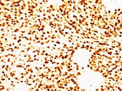
- Description: Recognizes a phosphor-protein of 45 kDa, identified as MyoD1. This MAb does not cross react with myogenin, Myf5, or Myf6. Antibody to MyoD1 labels the nuclei of myoblasts in developing muscle tissues. MyoD1 is not detected in normal adult tissue, but is highly expressed in the tumor cell nuclei of rhabdomyosarcomas. Occasionally nuclear expression of MyoD1 is seen in ectomesenchymoma and a subset of Wilm s tumors. Weak cytoplasmic staining is observed in several non-muscle tissues, including glandular epithelium and also in rhabdomyosarcomas, neuroblastomas, Ewing s sarcomas and alveolar soft part sarcomas._x000D__x000D_Primary antibodies are available purified, or with a selection of fluorescent CF® Dyes and other labels. CF® Dyes offer exceptional brightness and photostability. Note: Conjugates of blue fluorescent dyes like CF®405S and CF®405M are not recommended for detecting low abundance targets, because blue dyes have lower fluorescence and can give higher non-specific background than other dye colors.
- Synonyms: bHLHc1, Class C basic helix-loop-helix protein 1, Myoblast determination protein 1, Myogenic differentiation 1, Myogenic factor 3 (Myf-3), Myogenin D1, PUM
- CAS Number: 9007-83-4
- UNSPSC: 41116161
- UNSPSC Description: Primary and secondary antibodies for multiple methodology immunostaining detection application
- Gene Name: MYOD1
- Gene ID: 4654
- NCBI Gene ID: 181768
- UniProt: P15172
- Cellular Locus: Nucleus
- Host: Mouse
- Species Reactivity: Human
- Immunogen: Recombinant human MyoD1 protein
- Target Antigen: MyoD1
- Clonality: Monoclonal
- Isotype: IgG1 κ
- Clone: MYD712
- Conjugation: CF488A
- Disease: Tumor
- Source: Animal
- Field of Research: Cancer, Developmental biology
- Positive Control: Rhabdomyosarcoma
- Concentration: 0.1 mg/mL
- Buffer: PBS, 0.1% BSA, 0.05% azide
- Molecular Weight: 45 kDa
- Additionnal Information: For coating for ELISA, order Ab without BSA|Higher concentration may be required for direct detection using primary antibody conjugates than for indirect detection with secondary antibody|Optimal dilution and staining procedure for a specific application should be determined by user|Recommended starting concentrations for titration are 1-2 ug/mL for most applications, or 1 ug/million cells/100 uL for flow cytometry|Only nuclear staining should be considered as evidence of skeletal muscle differentiation
- References & Citations: Thulasi R et. al. Cell Growth and Differentiation, 1996, 7(4):531-41. | Wesche WA et. al. American Journal of Surgical Pathology, 1995, 19(3):261-9. | Parham DM et. al. Acta Neuropathologica, 1994, 87:605-11. |
- Shipping Conditions: Room temperature
- Storage Conditions: 4°C; Protect from light; Stable at room temperature or 37°C (98°F) for 7 days.
- Shelf Life: 2 years