
Anti-CD56 / NCAM(123C3.D5), CF488A conjugate
CAT:
37-BNC880015-100
Size:
100 µL
Price:
Ask
- Availability: 24/48H Stock Items & 2 to 6 Weeks non Stock Items.
- Dry Ice Shipment: No


Anti-CD56 / NCAM(123C3.D5), CF488A conjugate
- Description: This MAb reacts with an extracellular domain (close to transmembrane) of CD56/NCAM. Three isoforms of neural cell adhesion molecule (NCAM) are produced by differential splicing of the RNA transcript from a single gene. The 135 kDa isoform is the basic molecule, which is glycosylated or sialylated to produce the mature species. Anti-CD56 recognizes two proteins of the neural cell adhesion molecule, the basic molecule expressed on most neuroectodermally derived tissues and neoplasms (e.g. retinoblastoma, medulloblastomas, astrocytomas, neuroblastomas, and small cell carcinomas). It is also expressed on some mesodermally derived tumors (rhabdomyosarcoma). Anti-CD56 plays an important role in the diagnosis of nodal and nasal NK/T-cell lymphomas._x000D_ _x000D_ Primary antibodies are available purified, or with a selection of fluorescent CF® Dyes and other labels. CF® Dyes offer exceptional brightness and photostability. Note: Conjugates of blue fluorescent dyes like CF®405S and CF®405M are not recommended for detecting low abundance targets, because blue dyes have lower fluorescence and can give higher non-specific background than other dye colors._x000D_ _x000D_
- Synonyms: NCAM, Leu-19, NKH1, MSK39, NCAM120, NCAM140, NCAM180, Neural Cell Adhesion Molecule
- CAS Number: 9007-83-4
- UNSPSC: 41116161
- UNSPSC Description: Primary and secondary antibodies for multiple methodology immunostaining detection application
- Gene Name: NCAM1
- Gene ID: 4684
- NCBI Gene ID: 503878
- UniProt: P13591; P13592
- Cellular Locus: Plasma membrane
- Host: Mouse
- Species Reactivity: Human
- Immunogen: Membrane preparation of a small cell lung carcinoma
- Target Antigen: CD56 | NCAM
- Clonality: Monoclonal
- Isotype: IgG1 κ
- Clone: 123C3.D5
- Conjugation: CF488A
- Disease: Tumor
- Source: Animal
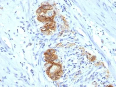
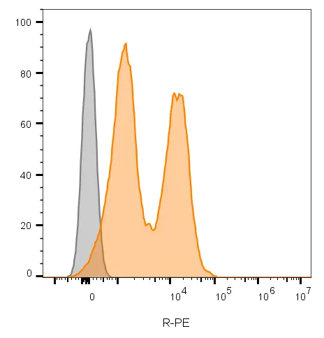
- Applications: Flow, surface (verified) | IF (verified) | IHC, FFPE (verified)
- Validated Applications: FC, IF, IHC, FFPE
- Field of Research: Cancer, Cell adhesion, Neuroscience
- Positive Control: Cerebellum, Pancreas, Neuroblastoma
- Concentration: 0.1 mg/mL
- Buffer: PBS, 0.1% BSA, 0.05% azide
- Molecular Weight: 180, 145 and 125 kDa
- Additionnal Information: Higher concentration may be required for direct detection using primary antibody conjugates than for indirect detection with secondary antibody|Immunofluorescence: 0.5-1 ug/mL|Immunohistology formalin-fixed 0.5-1 ug/mL|Staining of formalin-fixed tissues requires boiling tissue sections in 10 mM citrate buffer, pH 6.0, for 10-20 min followed by cooling at RT for 20 minutes|Flow Cytometry 0.5-1 ug/million cells/0.1 mL|Optimal dilution for a specific application should be determined by user
- Shipping Conditions: Room temperature
- Storage Conditions: 4°C; Protect from light; Stable at room temperature or 37°C (98°F) for 7 days.
- Shelf Life: 2 years