
Anti-p53 Tumor Suppressor Protein(PAb240)
CAT:
37-BNUM1634-50
Size:
50 µL
Price:
Ask
- Availability: 24/48H Stock Items & 2 to 6 Weeks non Stock Items.
- Dry Ice Shipment: No


Anti-p53 Tumor Suppressor Protein(PAb240)
- Description: The specificity of this monoclonal antibody to its intended target was tested by HuProt™ Array, containing more than 19,000 full-length human proteins. PAb240 binds to the C-terminus (aa213-217) of both wild type and mutated p53 in denatured samples and FFPE sections, but is reported to selectively detect mutant p53 in native samples by immunoprecipitation and ELISA. Mutation and/or allelic loss of p53 is one of the causes of a variety of mesenchymal and epithelial tumors. If it occurs in the germ line, such tumors run in families. p53 Binds to a DNA consensus sequence, the p53 response element, and it regulates normal cell growth cycle events by activating transcription of genes, involved either in progression through the cycle, or causing arrest in G1 when the genome is damaged. In most transformed and tumor cells the concentration of p53 is increased 51000 fold over the minute concentrations (1000 molecules cell) in normal cells, principally due to the increased half-life (4 h) compared to that of the wild-type (20 min). p53 Localizes in the nucleus, but is detectable at the plasma membrane during mitosis and when certain mutations modulate cytoplasmic/nuclear distribution. p53 Is the most commonly mutated gene in spontaneously occurring human cancers. Mutations arise with an average frequency of 70% but incidence varies from zero in carcinoid lung tumors to 97% in primary melanomas. High concentrations of p53 protein are transiently expressed in human epidermis and superficial dermal fibroblasts following mild ultraviolet irradiation._x000D_ _x000D_ Primary antibodies are available purified, or with a selection of fluorescent CF® Dyes and other labels. CF® Dyes offer exceptional brightness and photostability. Note: Conjugates of blue fluorescent dyes like CF®405S and CF®405M are not recommended for detecting low abundance targets, because blue dyes have lower fluorescence and can give higher non-specific background than other dye colors._x000D_ _x000D_
- Synonyms: Antigen NY-CO-13; BCC7; Cellular Tumor Antigen p53; LFS1; TP53; Transformation Related Protein 53 (TRP53); Tumor Protein p53; Tumor Suppressor p53
- CAS Number: 9007-83-4
- UNSPSC: 41116161
- UNSPSC Description: Primary and secondary antibodies for multiple methodology immunostaining detection application
- Gene Name: TP53
- Gene ID: 7157
- NCBI Gene ID: 654481
- UniProt: P04637
- Cellular Locus: Nucleus
- Host: Mouse
- Species Reactivity: Cow, Dog, Hamster, Human, Monkey, Mouse, Rat
- Immunogen: Gel-Purified p53-beta-galactosidase fusion protein containing murine p53 from aa 14-389
- Target Antigen: p53 Tumor Suppressor Protein
- Clonality: Monoclonal
- Isotype: IgG1 κ
- Clone: PAb240
- Conjugation: Purified, BSA-free
- Disease: Tumor
- Source: Animal
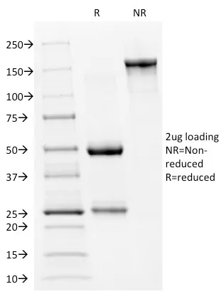
- Applications: IHC, FFPE (published) | IP (published) | WB (published)
- Validated Applications: IHC, FFPE, IP, WB
- Positive Control: MDA-MB-231 or A431 Cells. Breast or Colon carcinoma
- Concentration: 1 mg/mL
- Buffer: PBS, no BSA, no azide
- Molecular Weight: 53 kDa
- Additionnal Information: ELISA: For coating, order Ab without BSA; Optimal dilution for a specific application should be determined.|Higher concentration may be required for direct detection using primary antibody conjugates than for indirect detection with secondary antibody
- References & Citations: Note: References for this clone sold by other suppliers may be listed for expected applications. EMBO J (1990) 9(5): 1595-1602. (IP of mutant p53; IHC; WB) Oncogene (2003) 22: 4478-4487. (ELISA, mutant p53) Anticancer Res (2006) 26: 175-182. (IHC, FFPE)
- Shipping Conditions: Room temperature
- Storage Conditions: -35°C to -5°C ; Stable at room temperature or 37°C (98°F) for 7 days.
- Shelf Life: 2 years