
ERp57 (GRP58) Antibody
CAT:
400-SMC-168B
Size:
200 µg
Price:
Ask
- Availability: 24/48H Stock Items & 2 to 6 Weeks non Stock Items.
- Dry Ice Shipment: No


ERp57 (GRP58) Antibody
- Background: ERp57, also known as Glucose Regulated Protein 58 (Grp58), Hormone-Induced Protein-70 (HIP-70) and microsomal Carnitine Palmitoyltransferase, is a member of the protein disulfide isomerase family, containing two canonical CXHC tetrapeptide active site motifs (1-5). It has quite a few diverse roles. It functions as an accessory oxidoreductase involved in disulfide bond formation. In the ER, ERp57 interacts with membrane bound calnexin and soluble calreticulin (lectin chaperones) via their praline rich P-domain arms. Lectin chaperones bind nascent non-native glycoproteins, and position ERp57 to act upon the immature or misfolded glycoproteins that possess mono-glycosylated side chains. ERp57 deletion impairs posttranslational phases of influenza hema-glutinin folding, and causes accelerated release of MHC-I molecules, resulting in the coupling of sub-optimal peptides and reduced expression and stability on the cell surface (6). ERp57 also contains two thioredoxin active-site sequences, CGHC and an estrogen-binding domain. ERp57 is induced by both estrogen and leuteinizing-hormone-releasing hormone in the hippocampus (7).
- Description: Mouse Anti-Human ERp57 (GRP58) Monoclonal IgG1
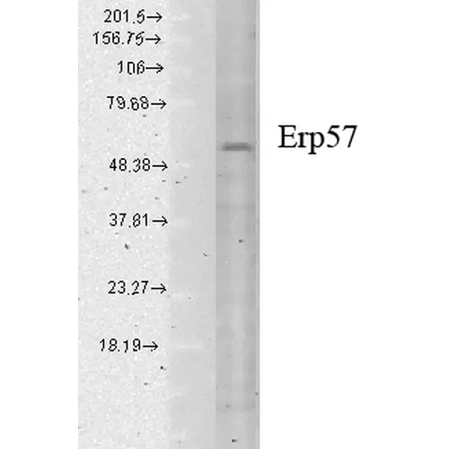
- Specifications: Detects ~57kDa.
- Product Name Alternative: ERp60 Antibody, ERp61 Antibody, Grp57 Antibody, Grp58 Antibody, P58 Antibody, PDIA3 Antibody, PI PLC Antibody, 58 kDa glucose regulated protein antibody, 58 kDa glucose-regulated protein antibody, 58 kDa microsomal protein antibody, Disulfide isomerase ER 60 antibody, Disulfide isomerase ER-60 antibody, Endoplasmic reticulum resident protein 57 antibody, Endoplasmic reticulum resident protein 60 antibody, ER p57 antibody, ER protein 57 antibody, ER protein 60 antibody, ERp 57 antibody, ERp57 antibody, Glucose Regulated Protein 58 Kd antibody, GRP 57 antibody, GRP 58 antibody, GRP57 antibody, HsT17083 antibody, p58 antibody, PDIA 3 antibody, PDIA3 antibody, PDIA3_HUMAN antibody, Phospholipase C alpha antibody, PI PLC antibody, Protein disulfide isomerase A3 antibody, Protein disulfide isomerase family A member 3 antibody, Protein disulfide-isomerase A3 antibody
- Synonyms: ERp57 (GRP58) Antibody, Clone Map.ERp57 (GRP58)
- CAS Number: 9007-83-4
- UNSPSC: 12352203
- Gene ID: 2923
- Swiss Prot: P30101
- Accession Number: NP_005304.3
- Cellular Locus: Endoplasmic Reticulum | Endoplasmic Reticulum Lumen | Melanosome
- Host: Mouse
- Species Reactivity: Human, Mouse, Rat, Bovine, Dog, Guinea Pig, Hamster, Monkey, Pig, Rabbit
- Immunogen: Human recombinant ERp57 (Grp58)
- Target: ERp57
- Clonality: Monoclonal
- Isotype: IgG1
- Clone: Map.ERp57
- Conjugation: Unconjugated
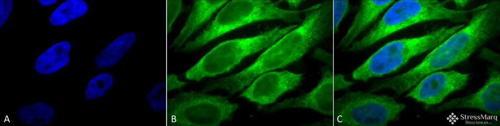
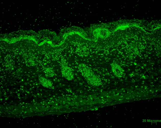
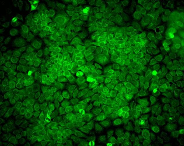
- Validated Applications: WB, IHC, ICC/IF, IP
- Purification: Protein G Purified
- Limit Of Detection: 0.5 µg/ml of SMC-168 was sufficient for detection of ERp57 in 10 µg of heat shock Heal Lysate by colorimetric immunoblot analysis using Goat anti-mouse IgG:HRP as the secondary antibody.
- Concentration: 1 mg/ml
- Dilution: WB (1:2000), IHC (1:100), ICC/IF (1:100); optimal dilutions for assays should be determined by the user.
- Weight: 0.2
- Buffer: PBS pH7.4, 50% glycerol, 0.09% sodium azide *Storage buffer changes when conjugated
- Precautions: Not for use in humans. Not for use in diagnostics or therapeutics. For in vitro research use only.
- References & Citations: 1. Herbert D.N. and Molinari M. (2007) Physiol Rev. 87: 1377-1408. 2. Williams D.B. (2005) J Cell Sci. 119: 615-623 3. Maattanen P., et al. (2006) Biochem Cell Biol. 84: 881-889. 4. Oliver J.D., et al. (1999) Mol Bio Cell. 10: 2573-2582. 5. Oliver J.D., et al. (1997) Science 275: 86-88. 6. Solda T., et al. (2006) J Biol Chem 281: 6219-6226. 7. Kimura T., et al. (2005) Biochem Biophys Research Communications. 331 (1): 224-230. 8. Chen, G., et al. (2002) Clin Cancer Res 8(7): 2298-2305. 9. Tan, P., et al. F. (2002) J Immunol 168(4): 1950-1960.