Anti-MyoD1 (Rhabdomyosarcoma Marker) (MYOD1/2075R), CF640R conjugate
CAT:
37-BNC402075-500
Size:
500 µL
Price:
Ask
- Availability: 24/48H Stock Items & 2 to 6 Weeks non Stock Items.
- Dry Ice Shipment: No


Anti-MyoD1 (Rhabdomyosarcoma Marker) (MYOD1/2075R), CF640R conjugate
Description:
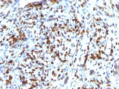
This antibody recognizes a phosphoprotein of 45 kDa, identified as MyoD1. This MAb does not cross react with myogenin, Myf5, or Myf6. Antibody to MyoD1 labels the nuclei of myoblasts in developing muscle tissues. MyoD1 is not detected in normal adult tissue, but is highly expressed in the tumor cell nuclei of rhabdomyosarcomas. Occasionally nuclear expression of MyoD1 is seen in ectomesenchymoma and a subset of Wilm's tumors. Weak cytoplasmic staining is observed in several non-muscle tissues, including glandular epithelium and also in rhabdomyosarcomas, neuroblastomas, Ewing s sarcomas and alveolar soft part sarcomas. Only nuclear staining should be considered as evidence of skeletal muscle differentiation.Primary antibodies are available purified, or with a selection of fluorescent CF® Dyes and other labels. CF® Dyes offer exceptional brightness and photostability. Note: Conjugates of blue fluorescent dyes like CF®405S and CF®405M are not recommended for detecting low abundance targets, because blue dyes have lower fluorescence and can give higher non-specific background than other dye colors.Synonyms:
bHLHc1, Class C basic helix-loop-helix protein 1, Myoblast determination protein 1, Myogenic differentiation 1, Myogenic factor 3 (Myf-3), Myogenin D1, PUMCAS Number:
9007-83-4UNSPSC:
41116161UNSPSC Description:
Primary and secondary antibodies for multiple methodology immunostaining detection applicationGene Name:
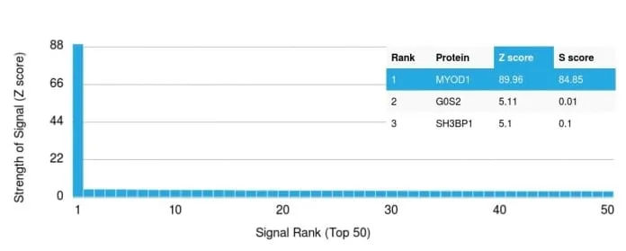
MYOD1Gene ID:
4654NCBI Gene ID:
181768UniProt:
P15172Cellular Locus:
NucleusHost:
RabbitSpecies Reactivity:
HumanImmunogen:
Recombinant full-length human MyoD1 proteinTarget Antigen:
MyoD1Clonality:
Recombinant MonoclonalIsotype:
IgGClone:
MYOD1/2075RConjugation:
CF640RDisease:
TumorSource:
AnimalApplications:
IHC, FFPE (verified)Validated Applications:
IHC, FFPEField of Research:
Cancer, Developmental biologyPositive Control:
Fetal skeletal muscle or RhabdomyosarcomaConcentration:
0.1 mg/mLBuffer:
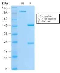
PBS, 0.1% BSA, 0.05% azideMolecular Weight:
45 kDaAdditionnal Information:
Higher concentration may be required for direct detection using primary antibody conjugates than for indirect detection with secondary antibody|Immunohistology (formalin): 1-2 ug/mL for 30 minutes at RT|Staining of formalin-fixed tissues requires boiling tissue sections in 10 mM Tris with 1 mM EDTA pH 9.0 for 10-20 minutes followed by cooling at RT for 20 minutes|Optimal dilution for a specific application should be determined by userShipping Conditions:
Room temperatureStorage Conditions:
4°C; Protect from light; Stable at room temperature or 37°C (98°F) for 7 days.Shelf Life:
2 years
DATASHEET Document
View DocumentMSDS Document
View Document