Anti-ZAP70(ZAP70/528), CF640R conjugate
CAT:
37-BNC400528-500
Size:
500 µL
Price:
Ask
- Availability: 24/48H Stock Items & 2 to 6 Weeks non Stock Items.
- Dry Ice Shipment: No


Anti-ZAP70(ZAP70/528), CF640R conjugate
Description:
ZAP70 is a 70 kDa protein tyrosine kinase found in T-cells and natural killer cells.Control of this protein translation is via the IgVH gene. ZAP70 protein is expressed in leukemic cells of approximately 25% of chronic lymphocytic leukemia (CLL) cases as well.Anti-ZAP70 expression is an excellent surrogate marker for the distinction between the Ig-mutated (anti-ZAP70 negative) and Ig-unmutated (anti-ZAP70 positive) CLL subtypes and can identify patient groups with divergent clinical courses. The anti-ZAP70 positive Ig-unmutated CLL cases have been shown to have a poorer prognosis.Primary antibodies are available purified, or with a selection of fluorescent CF® Dyes and other labels. CF® Dyes offer exceptional brightness and photostability. Note: Conjugates of blue fluorescent dyes like CF®405S and CF®405M are not recommended for detecting low abundance targets, because blue dyes have lower fluorescence and can give higher non-specific background than other dye colors.Synonyms:
Selective T cell defect; SRK; STD; Syk-related tyrosine kinase; Tyrosine-protein kinase ZAP-70;TZK; Zeta chain associated protein kinase 70kDaCAS Number:
9007-83-4UNSPSC:
41116161UNSPSC Description:
Primary and secondary antibodies for multiple methodology immunostaining detection applicationGene Name:
ZAP70Gene ID:
7535NCBI Gene ID:
234569UniProt:
P43403Cellular Locus:
CytoplasmicHost:
MouseSpecies Reactivity:
HumanImmunogen:
Recombinant full-length human ZAP70 proteinTarget Antigen:
ZAP70Clonality:
MonoclonalIsotype:
IgG2a κClone:
ZAP70/528Conjugation:
CF640RDisease:
TumorSource:
AnimalApplications:
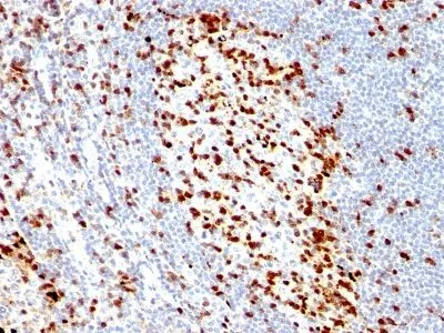
IHC, FFPE (verified)Validated Applications:
IHC, FFPEField of Research:
ImmunologyPositive Control:
Jurkat cells. Tonsil or lymph node.Concentration:
0.1 mg/mLBuffer:
PBS, 0.1% BSA, 0.05% azideMolecular Weight:
70 kDaAdditionnal Information:
Higher concentration may be required for direct detection using primary antibody conjugates than for indirect detection with secondary antibody|Immunofluorescence: 0.5-1 ug/mL|Immunohistology formalin-fixed 0.5-1 ug/mL|Staining of formalin-fixed tissues requires boiling tissue sections in 10 mM Tris with 1 mM EDTA, pH 9.0, for 10-20 min followed by cooling at RT for 20 minutes|Flow Cytometry 0.5-1 ug/million cells/0.1 mL|Optimal dilution for a specific application should be determined by userShipping Conditions:
Room temperatureStorage Conditions:
4°C; Protect from light; Stable at room temperature or 37°C (98°F) for 7 days.Shelf Life:
2 years
DATASHEET Document
View DocumentMSDS Document
View Document