Anti-CD7(T3-3A1), CF568 conjugate
CAT:
37-BNC680978-100
Size:
100 µL
Price:
Ask
- Availability: 24/48H Stock Items & 2 to 6 Weeks non Stock Items.
- Dry Ice Shipment: No


Anti-CD7(T3-3A1), CF568 conjugate
Description:
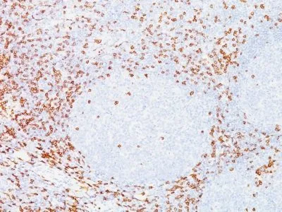
Recognizes a protein of 40 kDa, identified as CD7, a member of the immunoglobulin gene superfamily. Its N-terminal amino acids 1-107 are highly homologous to Ig kappa-L chains whereas the carboxyl-terminal region of the extracellular domain is proline-rich and has been postulated to form a stalk from which the Ig domain projects. CD7 is expressed on the majority of immature and mature T-lymphocytes, and T cell leukemia. It is also found on natural killer cells, a small subpopulation of normal B cells and on malignant B cells. Cross-linking surface CD7 positively modulates T cell and NK cell activity as measured by calcium fluxes, expression of adhesion molecules, cytokine secretion and proliferation. CD7 associates directly with phosphoinositol 3'-kinase. CD7 ligation induces production of D-3 phosphoinositides and tyrosine phosphorylation.Primary antibodies are available purified, or with a selection of fluorescent CF® Dyes and other labels. CF® Dyes offer exceptional brightness and photostability. Note: Conjugates of blue fluorescent dyes like CF®405S and CF®405M are not recommended for detecting low abundance targets, because blue dyes have lower fluorescence and can give higher non-specific background than other dye colors.Synonyms:
CD27 ligand (CD27L); CD27LG; Ki24; Surface antigen CD70; Tumor necrosis factor (ligand) superfamily, member 7 (TNFSF7)CAS Number:
9007-83-4UNSPSC:
41116161UNSPSC Description:
Primary and secondary antibodies for multiple methodology immunostaining detection applicationGene Name:
CD7Gene ID:
924NCBI Gene ID:
186820UniProt:
P09564Cellular Locus:
Plasma membraneHost:
MouseSpecies Reactivity:
HumanImmunogen:
Human T cellsTarget Antigen:
CD7Clonality:
MonoclonalIsotype:
IgG1 κClone:
T3-3A1Conjugation:
CF568Disease:
TumorSource:
AnimalApplications:
Flow, surface (published) | Functional studies (published) | IF (published) | ELISA (published) | IP (published)Validated Applications:
FC, IF, ELISA, IPField of Research:
Immunology, Signal transductionPositive Control:
Jurkat, HUT-78, Molt-4, CEM cells, or human peripheral blood lymphocytes. Lymph node and tonsil.Concentration:
0.1 mg/mLBuffer:
PBS, 0.1% BSA, 0.05% azideMolecular Weight:
40 kDaAdditionnal Information:
Higher concentration may be required for direct detection using primary antibody conjugates than for indirect detection with secondary antibody|Immunofluorescence: 0.5-1 ug/mL|Flow Cytometry 0.5-1 ug/million cells/0.1 mL|Optimal dilution for a specific application should be determined by userReferences & Citations:
Note: References for this clone sold by other suppliers may be listed for expected applications. PNAS USA (1979) 76: 5829-5833. PNAS USA (1980) 77(5): 2914-2918. (IP; functional studies) J Immunol Meth (1987) 96(2), 201-209. (cell ELISA) PNAS USA (1996) 93: 10348-10353. (cell depletion)>/li> Blood (1998) 91(8) 2760-2771. (Flow) J Cell Biol (1998) 142(5): 1245-1256. (IF; IP) Theranostics (2018) 8(21): 6070-6087. (mAb characterization; in vivo imaging)Shipping Conditions:
Room temperatureStorage Conditions:
4°C; Protect from light; Stable at room temperature or 37°C (98°F) for 7 days.Shelf Life:
2 years
DATASHEET Document
View DocumentMSDS Document
View Document