Anti-CD63(MX-49.129.5), CF488A conjugate
CAT:
37-BNC880525-500
Size:
500 µL
Price:
Ask
- Availability: 24/48H Stock Items & 2 to 6 Weeks non Stock Items.
- Dry Ice Shipment: No
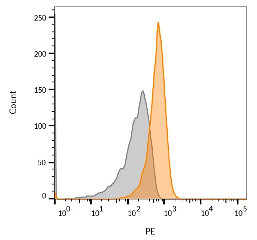
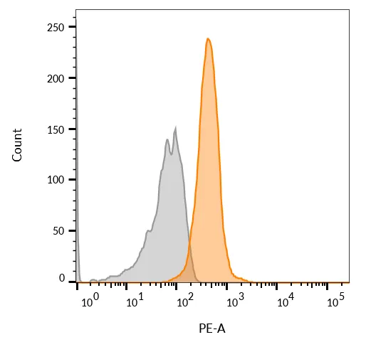
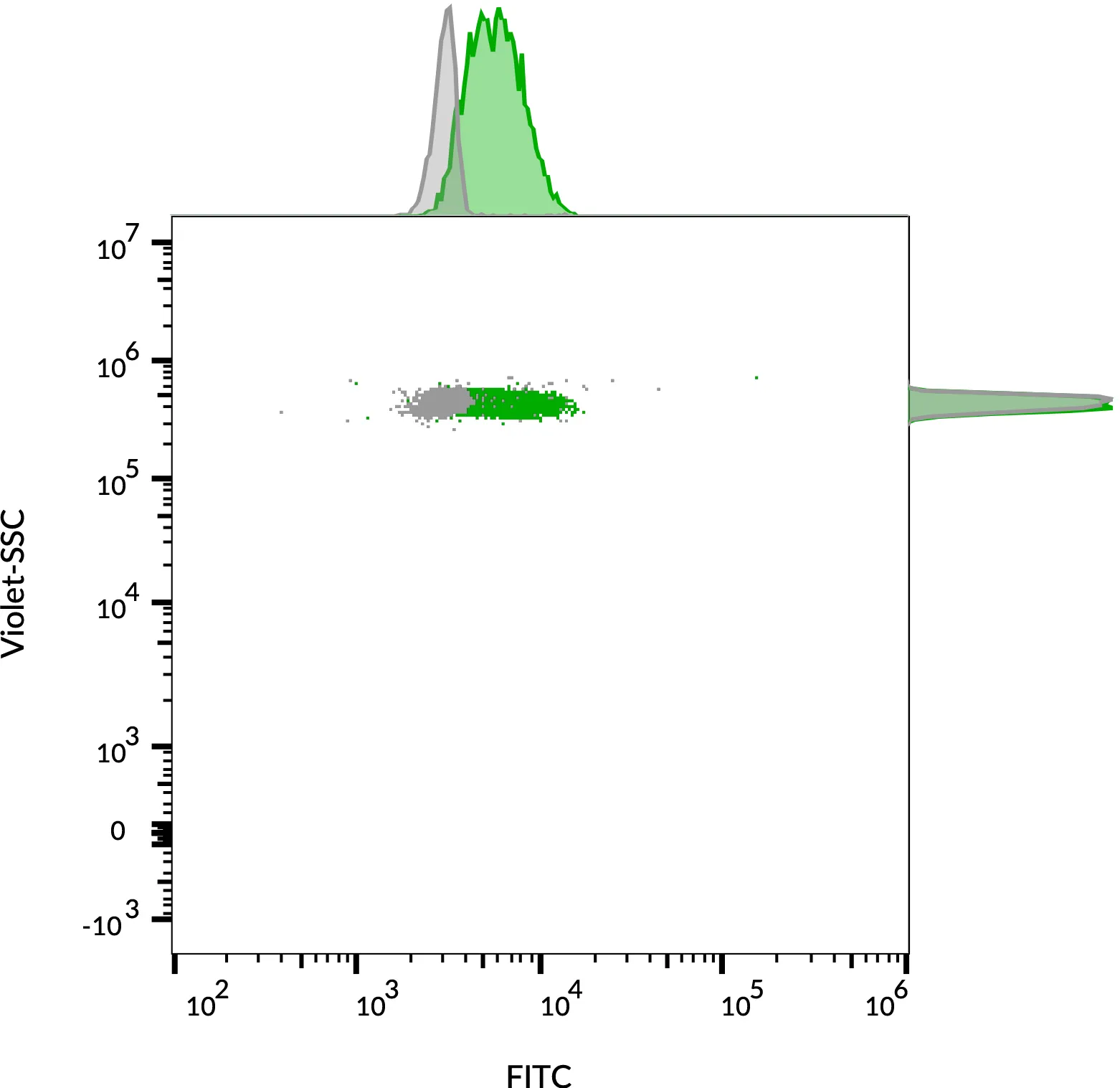
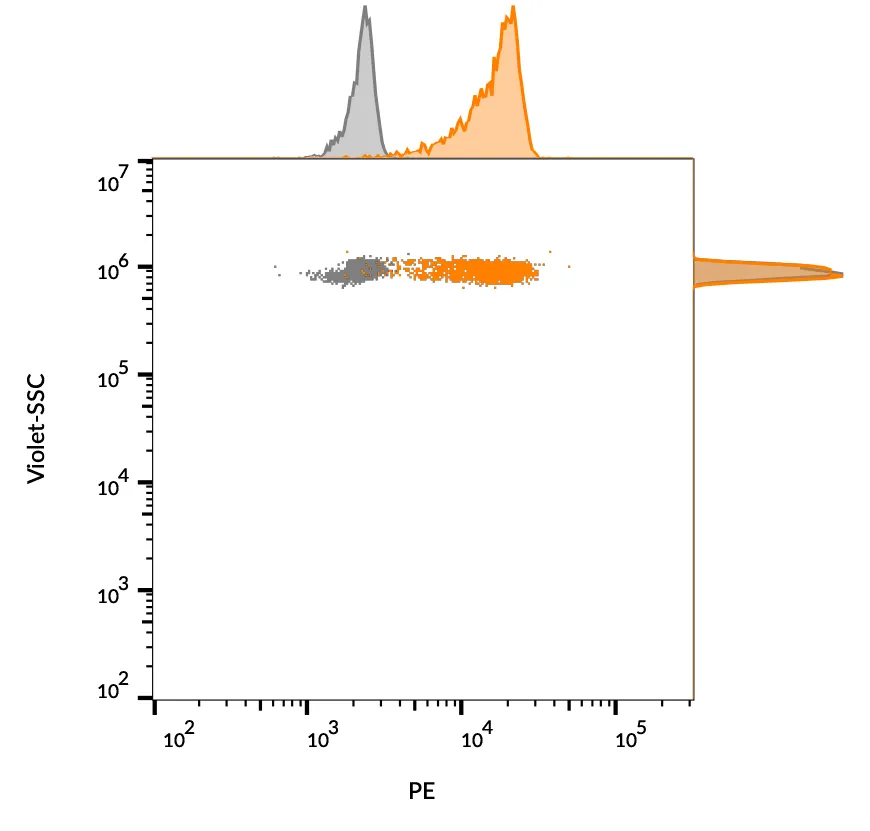


Anti-CD63(MX-49.129.5), CF488A conjugate
Description:
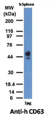
This MAb recognizes protein of 26 kDa-60 kDa, which is identified as CD63. Its epitope is different from that of MAb LAMP3/529. The tetraspanins are integral membrane proteins expressed on cell surface and granular membranes of hematopoietic cells and are components of multi-molecular complexes with specific integrins. The tetraspanin CD63 is a lysosomal membrane glycoprotein that translocates to the plasma membrane after platelet activation. CD63 is expressed on activated platelets, monocytes and macrophages, and is weakly expressed on granulocytes, T cell and B cells. It is located on the basophilic granule membranes and on the plasma membranes of lymphocytes and granulocytes. CD63 is a member of the TM4 superfamily of leukocyte glycoproteins that includes CD9, CD37 and CD53, which contain four transmembrane regions. CD63 may play a role in phagocytic and intracellular lysosome-phagosome fusion events. CD63 deficiency is associated with Hermansky-Pudlak syndrome and is strongly expressed during the early stages of melanoma progression._x000D_ _x000D_ Primary antibodies are available purified, or with a selection of fluorescent CF® Dyes and other labels. CF® Dyes offer exceptional brightness and photostability. Note: Conjugates of blue fluorescent dyes like CF®405S and CF®405M are not recommended for detecting low abundance targets, because blue dyes have lower fluorescence and can give higher non-specific background than other dye colors._x000D_ _x000D_Synonyms:
gp55; granulophysin; Lysosomal-associated membrane protein 3 (LAMP-3); Mast cell antigen AD1; melanoma 1 antigen; Melanoma-associated antigen MLA1; Melanoma-associated antigen ME491; MLA1; NGA; Ocular melanoma-associated antigen; OMA81H; PTLGP40; Tetraspanin-30; TSPAN30CAS Number:
9007-83-4UNSPSC:
41116161UNSPSC Description:
Primary and secondary antibodies for multiple methodology immunostaining detection applicationGene Name:
CD63Gene ID:
967NCBI Gene ID:
445570UniProt:
P08962Cellular Locus:
Plasma membraneHost:
MouseSpecies Reactivity:
Human, MouseImmunogen:
Full length CD63 of human originTarget Antigen:
CD63Clonality:
MonoclonalIsotype:
IgG1 κClone:
MX-49.129.5Conjugation:
CF488ASource:
AnimalApplications:
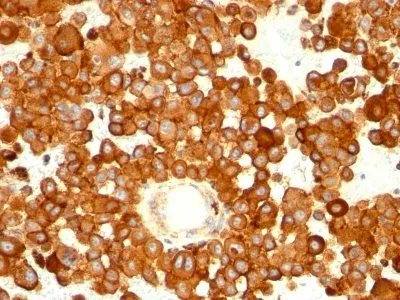
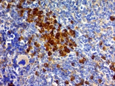
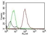
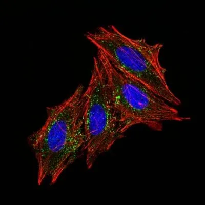
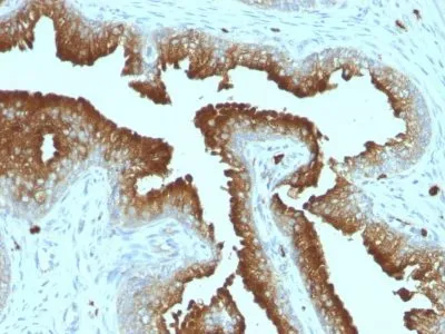
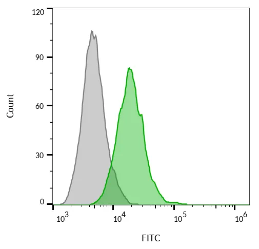
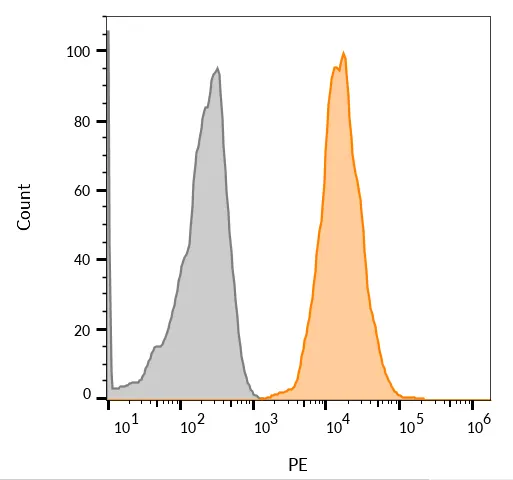
Exosome staining (verified) | Flow, surface (verified) | IF (verified) | IHC, FFPE (verified) | IP (published) | WB (verified)Validated Applications:
FC, IF, IHC, FFPE, IP, WBField of Research:
Cancer, Exosomes/EVs, ImmunologyPositive Control:
U87MG, SK-MEL-28, HL60, THP-1, NIH/3T3, or MCF-7 cells. Human melanoma, spleen or lymphoma tissue.Concentration:
0.1 mg/mLBuffer:
PBS, 0.1% BSA, 0.05% azideMolecular Weight:
26 kDa (core protein); 30-60 kDa (glycosylated)Additionnal Information:
Higher concentration may be required for direct detection using primary antibody conjugates than for indirect detection with secondary antibody|Immunofluorescence: 0.5-1 ug/mL|Immunohistology formalin-fixed 0.5-1 ug/mL|Staining of formalin-fixed tissues is enhanced by boiling tissue sections in 10 mM citrate buffer, pH 6.0, for 10-20 min followed by cooling at RT for 20 minutes|Flow Cytometry 0.5-1 ug/million cells/0.1 mL|Western blotting 0.5-1 ug/mL|Optimal dilution for a specific application should be determined by userReferences & Citations:
Note: References for this clone sold by other suppliers may be listed for expected applications. Mod Pathol (2010) 23: 1418-1428. (immunoprecipitation)Shipping Conditions:
Room temperatureStorage Conditions:
4°C; Protect from light; Stable at room temperature or 37°C (98°F) for 7 days.Shelf Life:
2 years
DATASHEET Document
View DocumentMSDS Document
View Document