Anti-CD35 / CR1(E11), CF640R conjugate
CAT:
37-BNC400040-100
Size:
100 µL
Price:
Ask
- Availability: 24/48H Stock Items & 2 to 6 Weeks non Stock Items.
- Dry Ice Shipment: No


Anti-CD35 / CR1(E11), CF640R conjugate
Description:
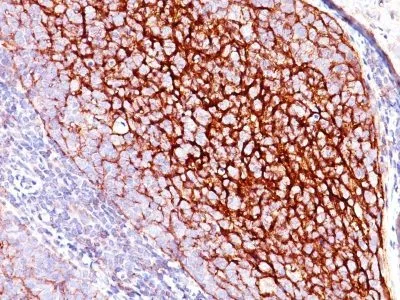
Recognizes a protein of 210-220 kDa, which is identified as the complement receptor 1 (CR1)/CD35. This MAb is specific for a site in CR1 that is distal from the C3b-binding site, so that it is unable to block CR1 activity. This MAb is highly specific to CR1 and shows no cross-reaction with CR2. The primary function of CR1 is to serve as the cellular receptor for C3b and C4b, the most important components of the complement system leading to clearance of foreign macromolecules. The Knops blood group system is a system of antigens located on this protein. Follicular dendritic cells (FDC) are restricted to the B-cell regions of secondary lymphoid follicles. They are CD21 /CD35 /CD1a-. This MAb labels follicular dendritic cells and follicular dendritic cell sarcoma.Primary antibodies are available purified, or with a selection of fluorescent CF® Dyes and other labels. CF® Dyes offer exceptional brightness and photostability. Note: Conjugates of blue fluorescent dyes like CF®405S and CF®405M are not recommended for detecting low abundance targets, because blue dyes have lower fluorescence and can give higher non-specific background than other dye colors.Synonyms:
C3 binding protein, C3b/C4b receptor, C3BR, C4BR, Complement Component (3b/4b) receptor 1 including Knops blood group system, Complement receptor type 1, KN, Knops blood group antigenCAS Number:
9007-83-4UNSPSC:
41116161UNSPSC Description:
Primary and secondary antibodies for multiple methodology immunostaining detection applicationGene Name:
CR1Gene ID:
1378NCBI Gene ID:
334019UniProt:
P17927Cellular Locus:
Plasma membraneHost:
MouseSpecies Reactivity:
Baboon, Cynomolgus, Human, Rhesus macaqueImmunogen:
Intact human monocytesTarget Antigen:
CD35 | CR1Clonality:
MonoclonalIsotype:
IgG1 κClone:
E11Conjugation:
CF640RSource:
AnimalApplications:
Flow, surface (published) | ELISA (published) | IHC, FFPE (verified) | WB (published)Validated Applications:
FC, ELISA, IHC, FFPE, WBField of Research:
ImmunologyPositive Control:
Follicular dendritic cells (FDC) in tonsilConcentration:
0.1 mg/mLBuffer:
PBS, 0.1% BSA, 0.05% azideMolecular Weight:
210-220 kDaAdditionnal Information:
Higher concentration may be required for direct detection using primary antibody conjugates than for indirect detection with secondary antibody|Immunofluorescence: 1-2 ug/mL|Immunohistology formalin-fixed 0.5-1 ug/mL|Staining of formalin-fixed tissues requires boiling tissue sections in 10 mM citrate buffer, pH 6.0, for 10-20 min followed by cooling at RT for 20 minutes|Flow Cytometry 0.5-1 ug/million cells/0.1 mL|Optimal dilution for a specific application should be determined by userReferences & Citations:
Note: References for this clone sold by other suppliers may be listed for expected applications. J Immunol (1999) 162:7549-7554. (WB) Clin Exp Immunol (2005) 143: 274-280. (ELISA) Int Immunol (2013) 25(1): 25-33. (Flow, surface)Shipping Conditions:
Room temperatureStorage Conditions:
4°C; Protect from light; Stable at room temperature or 37°C (98°F) for 7 days.Shelf Life:
2 years
DATASHEET Document
View DocumentMSDS Document
View Document