
Anti-Carbonic Anhydrase IX(CA9/781), Biotin conjugate
CAT:
37-BNCB0781-500
Size:
500 µL
Price:
Ask
- Availability: 24/48H Stock Items & 2 to 6 Weeks non Stock Items.
- Dry Ice Shipment: No


Anti-Carbonic Anhydrase IX(CA9/781), Biotin conjugate
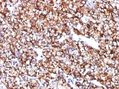
- Description: Carbonic anhydrase IX (carbonic anhydrase 9) is one of several carbolic anhydrases that vary in tissue distribution and localization. Carbonic anhydrases catalyze the interconversion of carbon dioxide and water into carbonic acid and bicarbonate and are found in all mammalian tissues. Carbonic anhydrase IX is a transmembrane protein that is not expressed in most healthy tissues, but is expressed in most renal cell carcinomas.Primary antibodies are available purified, or with a selection of fluorescent CF® Dyes and other labels. CF® Dyes offer exceptional brightness and photostability. Note: Conjugates of blue fluorescent dyes like CF®405S and CF®405M are not recommended for detecting low abundance targets, because blue dyes have lower fluorescence and can give higher non-specific background than other dye colors.
- Synonyms: Carbonic anhydrase IX; Carbonic anhydrase 9; RCC-associated antigen G250; Carbonic dehydratase; CA-IX; Membrane antigen MN
- CAS Number: 9007-83-4
- UNSPSC: 41116161
- UNSPSC Description: Primary and secondary antibodies for multiple methodology immunostaining detection application
- Gene Name: CA9
- Gene ID: 768
- NCBI Gene ID: 63287
- UniProt: Q16790
- Cellular Locus: Plasma membrane
- Host: Mouse
- Species Reactivity: Horse, Human
- Immunogen: Recombinant human CA9 protein
- Target Antigen: Carbonic Anhydrase IX
- Clonality: Monoclonal
- Isotype: IgG2b κ
- Clone: CA9/781
- Conjugation: Biotin
- Disease: Tumor
- Source: Animal
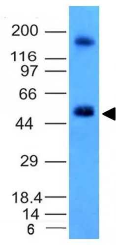
- Applications: IHC, FFPE (verified) | WB (verified)
- Validated Applications: IHC, FFPE, WB
- Field of Research: Cancer, Metabolism
- Positive Control: Normal kidney or renal cell carcinoma
- Concentration: 0.1 mg/mL
- Buffer: PBS, 0.1% BSA, 0.05% azide
- Molecular Weight: 55 kDa
- Additionnal Information: Higher concentration may be required for direct detection using primary antibody conjugates than for indirect detection with secondary antibody|Immunofluorescence: 1-2 ug/mL|Immunohistology formalin-fixed 0.5-1 ug/mL|Staining of formalin-fixed tissues requires boiling tissue sections in 10 mM citrate buffer, pH 6.0, for 10-20 min followed by cooling at RT for 20 minutes|Flow Cytometry 0.5-1 ug/million cells/0.1 mL|Western blotting 0.5-1 ug/mL|Optimal dilution for a specific application should be determined by user
- Shipping Conditions: Room temperature
- Storage Conditions: 4°C; Stable at room temperature or 37°C (98°F) for 7 days.
- Shelf Life: 2 years