Anti-CD3e (T-Cell Marker)(rC3e/1308), CF647 conjugate
CAT:
37-BNC473631-100
Size:
100 µL
Price:
Ask
- Availability: 24/48H Stock Items & 2 to 6 Weeks non Stock Items.
- Dry Ice Shipment: No


Anti-CD3e (T-Cell Marker)(rC3e/1308), CF647 conjugate
Description:
Recognizes the epsilon-chain of CD3, which consists of five different polypeptide chains (designated as gamma, delta, epsilon, zeta, and eta) with MW ranging from 16-28 kDa. The CD3 complex is closely associated at the lymphocyte cell surface with the T cell antigen receptor (TCR). Reportedly, CD3 complex is involved in signal transduction to the T cell interior following antigen recognition. The CD3 antigen is first detectable in early thymocytes and probably represents one of the earliest signs of commitment to the T cell lineage. In cortical thymocytes, CD3 is predominantly intra-cytoplasmic. However, in medullary thymocytes, it appears on the T cell surface. CD3 antigen is a highly specific marker for T cells, and is present in majority of T cell neoplasms._x000D_ _x000D_ Primary antibodies are available purified, or with a selection of fluorescent CF® Dyes and other labels. CF® Dyes offer exceptional brightness and photostability. Note: Conjugates of blue fluorescent dyes like CF®405S and CF®405M are not recommended for detecting low abundance targets, because blue dyes have lower fluorescence and can give higher non-specific background than other dye colors._x000D_ _x000D_Synonyms:
CD3 epsilon; CD3 TCR complex; CD3e antigen epsilon polypeptide (TiT3 complex); T-cell surface antigen T3/Leu-4 epsilon chain; T-cell surface glycoprotein CD3 epsilon chain; T3E; TCRE; TiT3 complexCAS Number:
9007-83-4UNSPSC:
41116161UNSPSC Description:
Primary and secondary antibodies for multiple methodology immunostaining detection applicationGene Name:
CD3EGene ID:
916NCBI Gene ID:
3003UniProt:
P07766Cellular Locus:
Plasma membraneHost:
MouseSpecies Reactivity:
HumanImmunogen:
Recombinant full-length human CD3E proteinTarget Antigen:
CD3eClonality:
Recombinant MonoclonalIsotype:
IgG1 κClone:
rC3e/1308Conjugation:
CF647Disease:
TumorSource:
AnimalApplications:
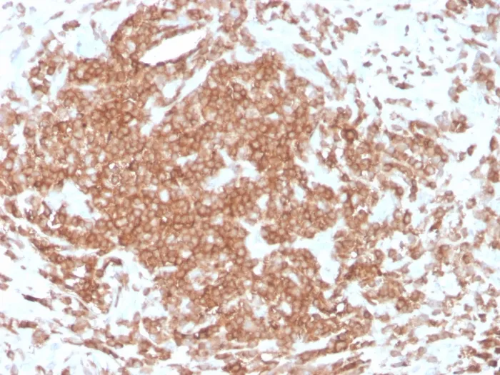
IHC, FFPE (verified)Validated Applications:
IHC, FFPEPositive Control:
Jurkat cells. Tonsil and lymph nodeConcentration:
0.1 mg/mLBuffer:
PBS, 0.1% BSA, 0.05% azideMolecular Weight:
20 kDaAdditionnal Information:
Not recommended for flow cytometry|Immunohistology (formalin) 1-2 ug/mL|Staining of formalin-fixed tissues requires boiling tissue sections in 10 mM citrate buffer, pH 6.0, for 10-20 min followed by cooling at RT for 20 min|Western blotting 1-2 ug/mL|Immunofluorescence 1-2 ug/mL|Optimal dilution for a specific application should be determined by userShipping Conditions:
Room temperatureStorage Conditions:
4°C; Protect from light; Stable at room temperature or 37°C (98°F) for 7 days.Shelf Life:
2 years
DATASHEET Document
View DocumentMSDS Document
View Document